
Figure 6.
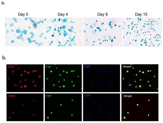
Analysis of β-galactosidase expression in LDPCs derived from double transgenic AlbCreXRosa26 mice. (A) X-gal staining of LDPC cultures on day 0 showed that nearly all hepatocytes were x-gal positive. On Day 4, dedifferentiating hepatocytes began to give rise to small x-gal positive cells which become more prominent by Day 8. On Day 15, virtually all cells in the culture were x-gal positive LDPCs (original magnification in all panels: 100x). (B) Confirmation of β-galactosidase positive cells as LDPCs. The round small cells that emerged in the cultures set up with hepatocytes from AlbCreXRosa26 mice were subjected to co-staining for β-galactosidase and LDPC markers CD45 and LMO2 (original magnification in all panels: 100x). The results showed complete overlap of the β-galactosidase and LDPC markers confirming that the cells were indeed LDPCs expressing β-galactosidase. These findings strongly support the hypothesis that LDPCs directly originate from hepatocytes.