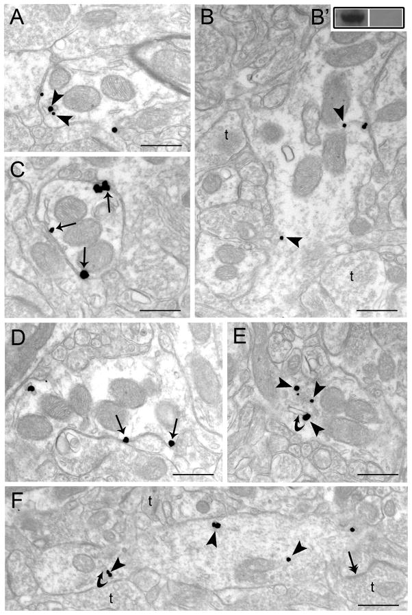
Figure 1.
Electron microscopic evidence for agonist-induced trafficking of WLS in rat striatum. Sections from control (vehicle-treated) (A, B), morphine-treated (C–D) and DAMGO-treated rats (E–F). A–B. Immunogold-silver labeling for WLS (arrowheads) can be seen in dendrites from vehicle-treated rats. B′. Western blot analysis showing WLS immunoreactivity in a frontal cortex microsample (left lane) and a preabsorption control using the immunizing protein (right lane). C–D. WLS labeling is more prominently distributed along the plasmalemma of striatal dendrites following morphine treatment. Arrows point to immunogold-silver labeling along the plasmalemma. E–F. Immunogold-silver labeling for WLS can be seen within the cytoplasm (arrowheads) in dendrites from a DAMGO-treated rat. Curved arrows point to endosome-like vesicles. Double headed arrows indicate synaptic specializations formed by axon terminals (t). Scale bars, 0.5μm.

