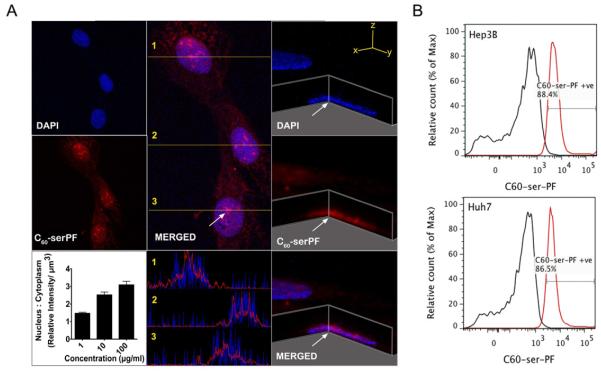
Figure 2. C60-serPF NPs localize to the nucleus liver cancer cells.
Panel A. Confocal microscopy image of Hep3B cells and Z-stack through the nucleus at the point depicted by the arrow. Intensity profiles through line 1, 2, and 3 are also represented, demonstrating colocalization with DAPI. Concentration of C60-serPF in the nucleus increases relative to cytoplasm as the cells are exposed to increasing concentrations of C60-serPF. Panel B. Nuclear uptake is confirmed by flow cytometry after extraction of nuclei demonstrating uptake in the majority of cells.

