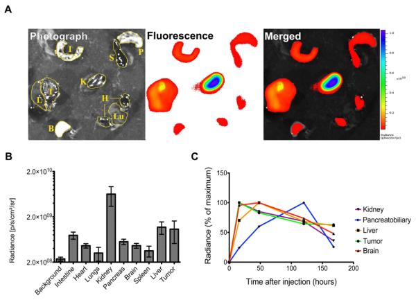
Figure 6. Relative biodistribution of C60-serPF in an animal model of primary liver cancer and in vivo nuclear localization.
Panel A. Fluorescence images from tumor (T), Liver (L), Intestine (I), Pancreatobiliary (P), Spleen (S), Kidney (K), Brain (B), bisected heart (H), and Lungs (Lu) of a mouse bearing an orthotopic liver tumor generated after injecting Hep3B cells into the liver. Panel B. Relative tissue distribution of C60-serPF in mouse tissues, demonstrating accumulation in most tissues at 16-hours and predominantly in the kidney, liver, and liver cancer. (n=3) Panel C. Pharmacokinetics of fullerene uptake in select tissues, suggesting early renal clearance and a delayed hepatic clearance. C60-serPF localizes to the brain and tumor within 16 hours and persists for more than one week (one mouse per time point represented).

