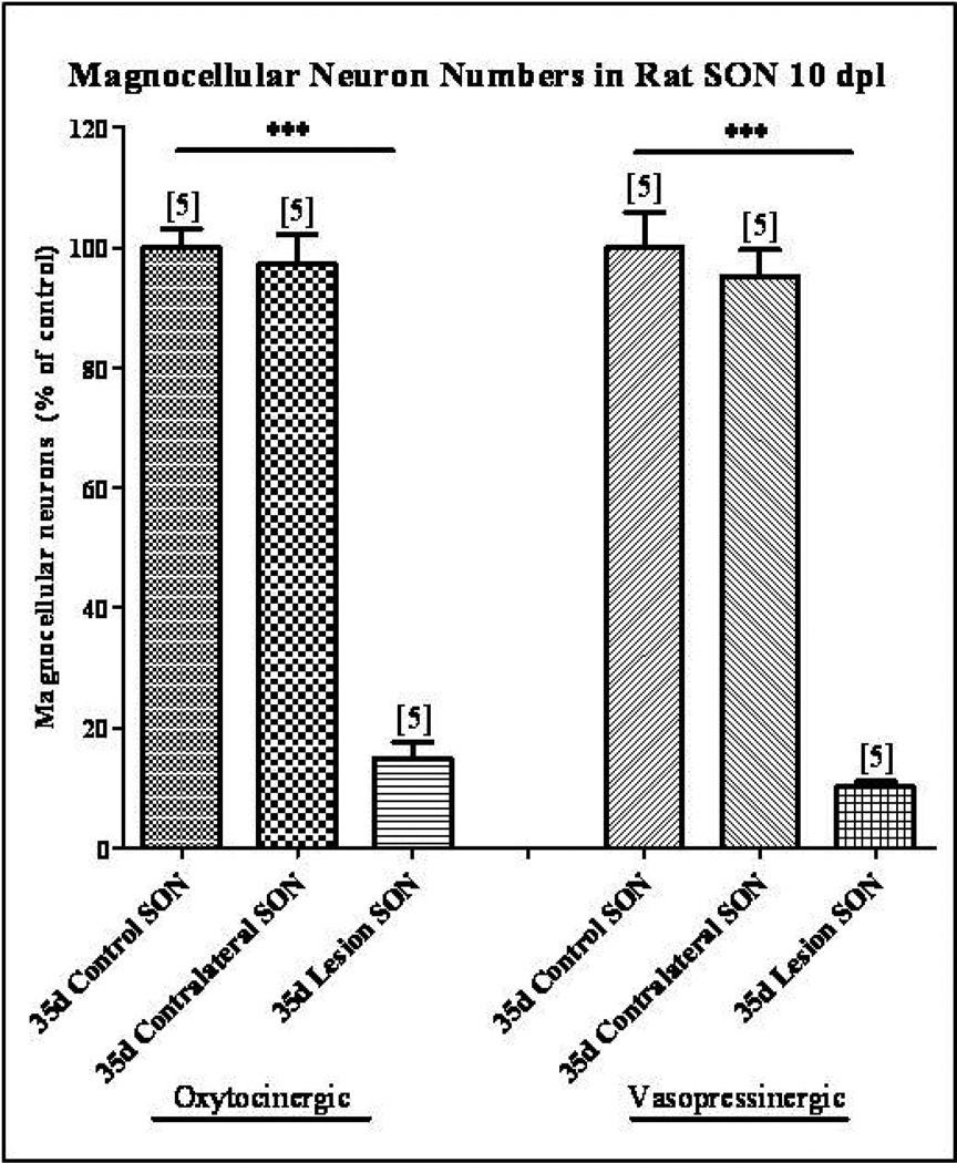
Fig. 2. Magnocellular neuron survival following unilateral lesion.
Immunohistochemical labeling of OT and VP neurons was used to identify individual magnocellular neurons. Cell counts demonstrated no significant decrease in the number of OT or VP neurons in the SON contralateral to the unilateral lesion. However, at 10 dpl the numbers of OT and VP neurons were reduced by 85% and 90% respectively in the axotomized SON. Column bars and error bars represent the mean and SEM of each group. Each group is comprised of a minimum of six sections sampled from each of 5 animals. ***p<0.0001.

