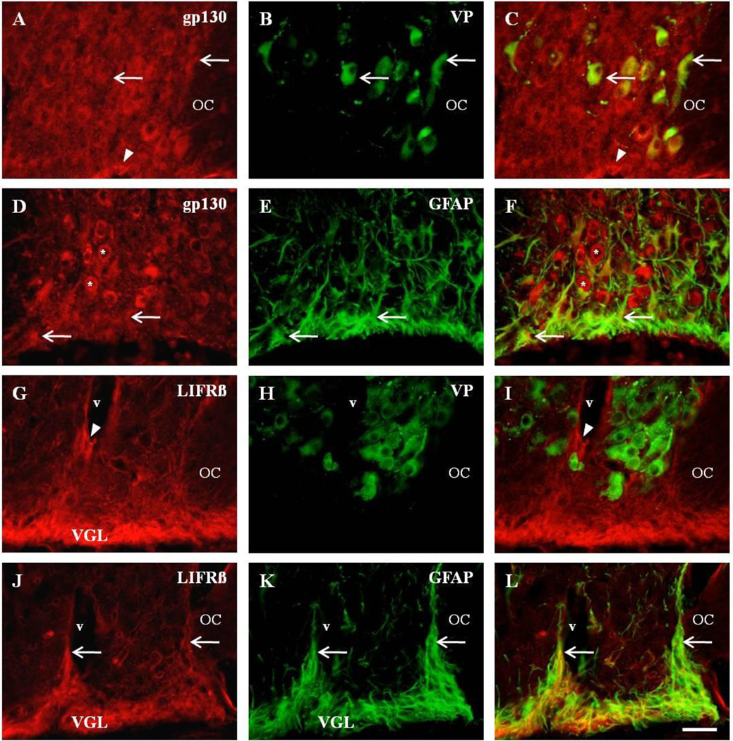
Fig. 6. Differential expression of CNTF receptor complex on astrocytes and magnocellular neurons.
Dual fluorescent colocalization of anti-gp130 (A), with anti-VP (B), revealed colocalization in vasopressinergic neurons (C, arrows). Note the gp130-immunoreactive profiles in presumptive astrocytes in the ventral glial limitans (VGL) of the SON (A, C, arrowheads). Similar observations were observed with anti-OT (not shown). Immunocytochemical analysis also demonstrated colocalization of anti-gp130 (D), with GFAP-immunoreactive astrocytes (E), of the SON (F, arrows). Also present are presumptive magnocellular neurons that are immunopositive for gp130 (D, F, asterisks). Unlike gp130, there was no observable colocalization of anti-LIFRβ in the magnocellular neurons of the SON (G–I). Note the LIFRβ-immunoreactive profiles surrounding blood vessels (v) in the SON (G, I, arrowheads) which do not colocalize with anti-VP (H) or anti-OT (not shown). However, strong LIFRβ-immunoreactivity in the ventral glial limitans (VGL) of the SON (J), revealed extensive colocalization of anti-LIFRβ (J), with anti-GFAP (K), throughout the entire SON (L, arrows). OC, optic chiasm; OT, oxytocin; v, blood vessel; VGL, ventral glial limitans; VP, vasopressin. Scale bar = 50 µm.

