
Figure 4.
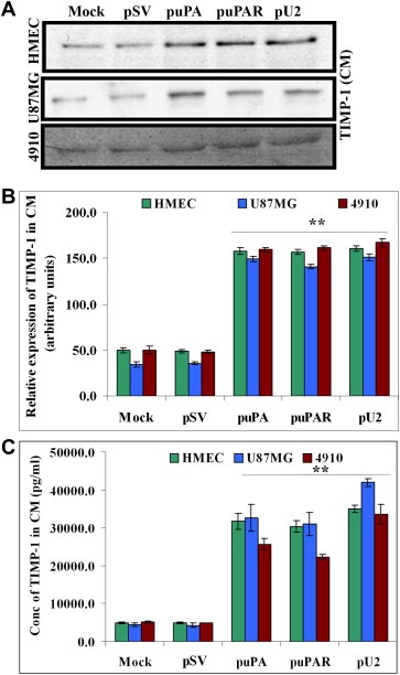
shRNA against uPA and uPAR enhances secretion of TIMP‐1 from HMEC, U87MG and 4910 cells by Western Blot and ELISA. (A) (Upper panel) 1.5×10⁵ HMEC, U87MG and 4910 cells were transfected as described earlier. Conditioned medium was collected after 72h and TIMP‐1 levels determined by Western blotting. Equal amount of conditioned medium was loaded after normalization of protein content. (B) Graph represents quantification of Western blotting results using NIH Image J software. (C) 1.5×10⁵ HMEC, U87MG and 4910 cells were transfected as described earlier. Conditioned medium was collected after 72h and TIMP‐1 levels determined by ELISA as per the manufacturer's instructions. Data represented are the average of three independent experiments. ∗p<0.01; ∗∗p<0.001.