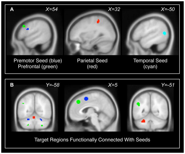
Figure 1. Regions of interest.
(A) Seed regions were areas where patients exhibited fMRI responses to finger tapping but controls did not. Seed regions were located in right premotor cortex (blue), right prefrontal cortex (green), right parietal cortex (red), and left temporal cortex (cyan). Seed regions are further described in Table 2. (B) Additional target regions of interest in cerebellum, preSMA, medial frontal cortex, and left parietal lobe were defined from the connectivity maps of the seeds, using voxels that exhibited significant intrinsic functional connectivity during the finger-tapping experiment (including both patients and controls, p<0.001 uncorrected). These are color-coded according to the seed region that produced them. The target regions for the middle temporal seed are not shown; they overlap with the target regions for the premotor seed. Target regions are further described in Table 4.

