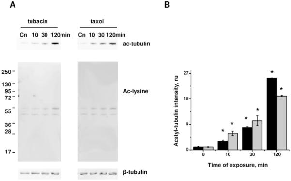
Figure 6.
Effect of tubacin and taxol on tubulin acetylation. HPAEC monolayers treated with 5 μM tubacin or 10 μM taxol for the time indicated were extracted and analyzed by Western blot with anti-acetyl-tubulin and anti-acetylated lysine antibodies (A). Beta-tubulin staining was used as a loading control. B) Normalized aceto-tubulin intensities (black columns, tubacin; grey columns, taxol) were expressed as fold of control. Shown are mean±SEM, *p<0.05 are considered significantly different.

