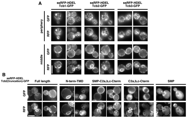
Fig. 3.
The tricalbins localize to ER–PM contacts. (A) Cells expressing Tcb1–GFP, Tcb2–GFP or Tcb3–GFP and the ER marker RFP–HDEL (strains WPY971, WPY973 and WPY975, respectively). Images were taken focusing on the middle and periphery of the cells. (B) Localization of GFP fused to the indicated portions of Tcb2p. The fusion proteins, under the GALl1 promoter, were expressed for 2 hours in strains containing the ER marker RFP–HDEL (strains ATY33, ATY34, ATY36, ATY37, ATY38). Scale bars: 5 μm.

