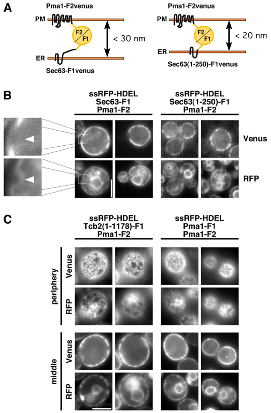
Fig. 4.
The cortical ER is closely apposed to the plasma membrane. (A) Diagram of the interaction between Sec63-F1 in the ER and Pma1-F2 in the PM. Sec63(1–250)-F1 contains the first 250 amino acids of Sec63p and lacks the C-terminal cytosolic domain of Sec63p. (B,C) Cells expressing the indicated fusion proteins and the ER marker RFP–HDEL (strains ATY57, ATY98, ATY97, ATY54). Arrowheads indicate peripheral ER that is not close to the PM and lacks Venus signal. The images in C were taken focusing on the middle and periphery of the cells. Scale bars: 5 μm.

