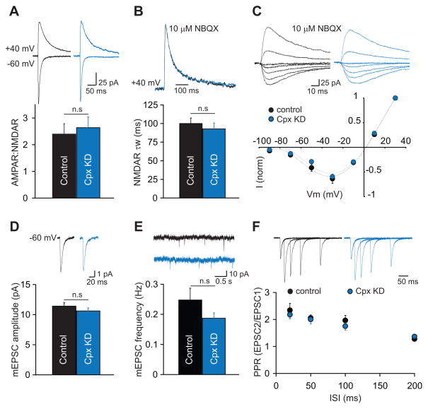
Figure 2. Postsynaptic Knockdown of Complexin-1 and -2 (Cpx KD) In Vivo Does Not Alter Basal Synaptic Transmission.
(A) The ratio of AMPAR- to NMDAR-mediated EPSCs is unchanged in Cpx KD cells. Representative EPSCs recorded at −60 mV and +40 mV are shown above the bar graph. (B) The weighted decay time constant of isolated NMDAR EPSCs at +40 mV is unchanged by postsynaptic Cpx KD. Scaled NMDAR EPSCs from two representative cells are shown above the bar graph. (C) Postsynaptic Cpx KD does not affect the I/V relationship of isolated NMDAR EPSCs. Representative traces from the two cells are shown above the graph. (D) The mean amplitude of mEPSCs is unaltered by postsynaptic Cpx KD. Averaged mEPSCs from representative cells are shown above the graph. (E) The mean frequency of mEPSCs is also unchanged by Cpx KD. Short traces of recordings from representative cells are shown above the graph. (F) Paired-pulse ratios of AMPAR EPSCs are unchanged in Cpx KD cells. EPSCs from two representative cells are shown above the graph. In all panels, bar graphs and individual points represent means ± SEM.

