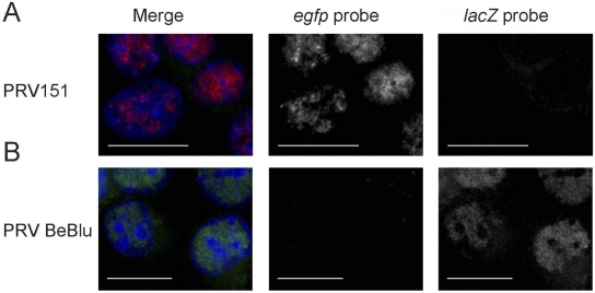
FIG 4 .
Similar viral replication centers after infection with two PRV recombinants. PK15 cells were infected by PRV151 (A) or PRV BaBlu (B) at an MOI of 10. The cells were then fixed at 4 hpi and hybridized with an egfp probe labeled with Alexa Fluor 647 (labeled in red) and a lacZ probe labeled with Alexa Fluor 488 (labeled in green). DAPI-labeled nuclei are in blue. Images are a maximal projection of 5 slices (0.5 µm apart) from a confocal microscope. Scale bar, 20 µm.

