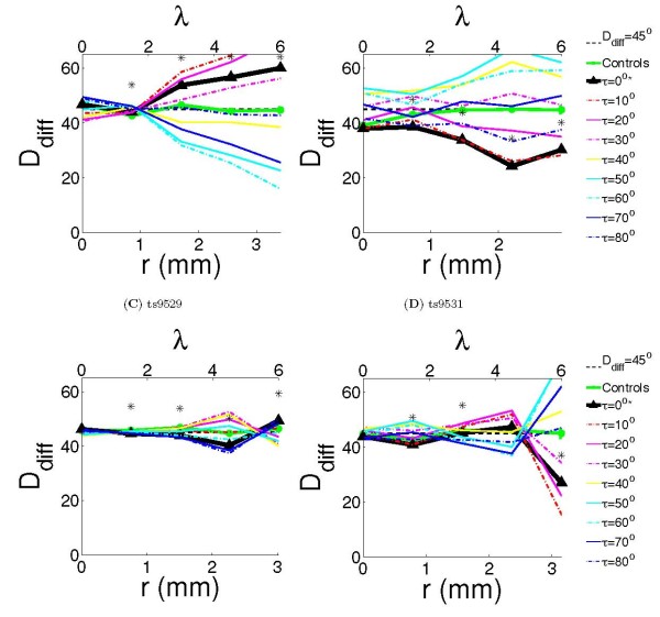
Figure 4.
Quantification of co-circularity. Ddiff was calculated for all cases as a function of cortical distance, indicated in both millimeters and wavelengths λ. The dotted black line marks Ddiff(r) = 45°, deviations below this line indicate co-circularity. Significance (P ≤ 0.05) compared with the control cases is marked as '*' on figure. We also report  (r) and the difference Ddiff(r, 0) − Ddiff(r,
  (r)). (A) Case ts9509 had Ddiff significantly different from the control case at r = 1.5, 3, 4.5, 6λ (P <0.01). The values of τ which minimized the Ddiff at r = 1.5, 3, 4.5, 6λ were 10°, 60°, 60°, 60° respectively, with differences Ddiff(r, 0) − Ddiff(r,
  (r)) of 0.2°, 21.9°, 31.2°, 44.0° respectively. (B) Case ts9514 had Ddiff significantly different from the control case at r = 1.5, 3, 4.5, 6λ (P <0.01). The values of τ which minimized the Ddiff at r = 1.5, 3, 4.5, 6λ were 0°, 10°, 0°, 10° respectively, with differences Ddiff(r, 0) − Ddiff(r,
  >(r)) of 0°, 0.08°, 0°, 2° respectively. (C) Case ts9529 had Ddiff significantly different from the control case at r = 1.5, 3, 4.5, 6λ (P <0.01). The values of τ which minimized the Ddiff at r = 1.5, 3, 4.5, 6λ were 0°, 80°, 80°, 40° respectively, with differences Ddiff(r, 0) − Ddiff(r,
  (r)) of 0°, 0.7°, 2.9°, 9.3° respectively. (D) Case ts9531 had Ddiff significantly different from the control case at r = 1.5, 3, 6λ (P <0.04). The values of τ which minimized the Ddiff at r = 1.5, 3, 4.5, 6λ were 0°, 70°, 60°, 10° respectively, with differences Ddiff(r, 0) − Ddiff(r,
  (r)) of 0°, 3.9°, 10.3°, 11.5° respectively.

