
Figure 11. Engrailed-GFP as a reporter for teratogenesis.
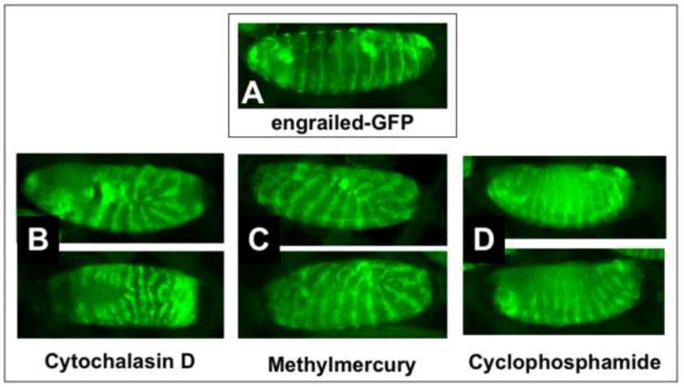
A) An engrailed-GFP embryo exhibiting normal development after EPS permeabilization at approximately 2-4 hours (EPS 1:10, one minute; Rhodamine B 1mM, 5min.). Abnormal development is seen in similarly permeabilized engrailed-GFP embryos incubated in cytochalasin D (50μM, B), methylmercury (50μM, C) and cyclophosphamide (100μM, E) and developed for 20 hours at 25°C. Images captured with a Nikon SMZ1500 stereo dissecting microscope. Anterior is left and dorsal is up (except in the lower panel of B which is a ventral view. See text for discussion).