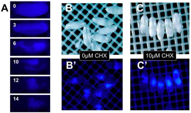
Figure 8. An intrinsic fluorescent indicator of embryo development.

Blue auto-fluorescence can be detected in the yolk proteins. Change in the distribution pattern of blue fluorescence over 14 hours of development (stage 5-14, panel A) is seen in representative frames of time-lapse imaging. Blue fluorescence in the midgut is easily discernable in normally developed EPS-permeablized (1:10 EPS one minute) Stage 16/17 embryos (Panel B, B’). Permeabilized embryos exposed to 10 μM cycloheximide (CHX) shows gut malformation as seen by the blue fluorescence centrally localized in a “ball” of tissue or dispersed anteriorly and posteriorly (Panel C, C’. See text for discussion).
