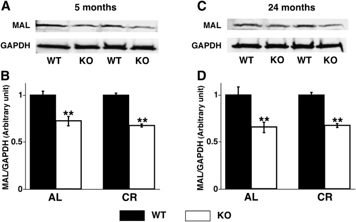
Fig. 4.
Cortical expression of myelin and lymphocyte protein was decreased in PGC-1α KO mice. Expression levels of MAL in PGC-1α KO compared with WT mice under AL and CR at 5 months (A, B) and 24 months (C, D) of age were analyzed by Western blot analysis (see Materials and Methods). Data represent mean ± SE of four animals per group after normalization to loading control GAPDH. Statistical significance was determined by a two-tailed Student t-test; **P < 0.01.

