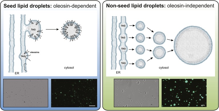
Fig. 2.
Schematic representation of models for oleosin-dependent and oleosin-independent LD formation from the ER in plant cells. In seed tissues (left panel), oleosins are cotranslationally inserted into the ER where they partition into domains in which TAG is accumulating between two leaflets. This promotes orientation of the oleosin proteins with N- and C termini facing the cytosol and the rest of the hydrophobic region of the protein adopting an extended hairpin configuration in the TAG matrix. The LDs in the cytosol are stabilized by oleosins and kept from fusing despite rapid dehydration and rehydration of these tissues during seed desiccation and imbibition. Micrographs are of isolated LDs from Arabidopsis seeds in bright-field (left) or by epifluorescence (right) following staining with Bodipy 493/503, a neutral lipid selective stain. LDs in nonseed tissues (right panel) may form from smaller TAG droplets that initially pinch off from the ER, then fuse to form larger droplets. Oleosins are not present in these LDs and the protein composition of LDs in nonseed tissues remains unknown. Micrographs are of LDs isolated from the oleaginous mesocarp of avocado fruit imaged in bright-field (left) or by Bodipy493/503 fluorescence (right). The white bars represent 50 microns. LDs from seeds tend to be smaller and more uniform compared with those from nonseed tissues, which fuse readily even in solution. Figure prepared by Dr. Charlene Case and Mr. Patrick Horn.

