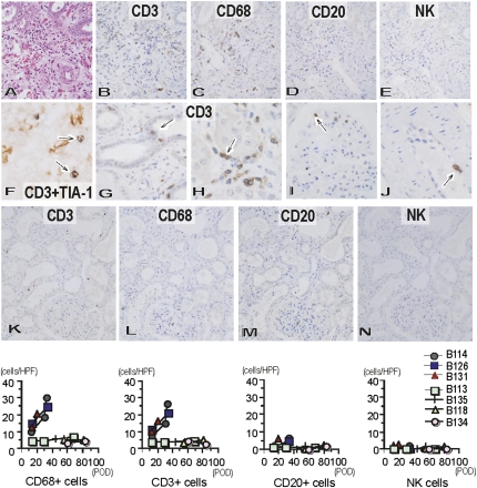
Figure 7.
Cellular infiltration within Gal knockout kidneys in baboons treated with chronic immunosuppression or immunotolerance protocol. Cellular infiltration in immunosuppression (A–J: B114, postoperative day 34) and immunotolerance (K–N: B134, postoperative day 83) protocols. Cellular infiltration developed at postoperative day 34 (B114) (A: hematoxylin and eosin stain; original magnification, ×400) in the immunosuppression protocol with infiltration of CD3+ T cells (B: original magnification, ×400) and CD68+ macrophages (C: original magnification, ×400), although with little infiltration of CD20+ B cells (D: original magnification, ×400) and NK cells (E: original magnification, ×400). Many CD3+ (brown) T cells contained TIA-1+ (blue) cytotoxic granules (arrow) (F: original magnification, ×900), capillaritis, acute glomerulitis, and endothelialitis developed with CD3+ T cells (arrow) in tubules (G: original magnification, ×800), peritubular capillaries (H: original magnification, ×800), glomeruli (I: original magnification, ×800), and arteries (J: original magnification, ×800). In contrast, only small numbers of CD3+ T cells (K: original magnification, ×200) and CD68+ macrophages (L: original magnification, ×200), and very few CD20+ B cells (M: original magnification, ×200) and NK cells (N: original magnification, ×200) were present in the grafts at postoperative day 83 in B134 in the immunotolerance protocol. Analysis of the number of infiltrating CD3+ T cells, macrophages, CD20+ B cells, and NK cells in the grafts (cells/single field of 0.0625 mm2) indicated increased infiltration of T cells and macrophages in the grafts of the immunosuppression protocol, but not those of the immunotolerance protocol. HPF, high-power field; POD, postoperative day.

