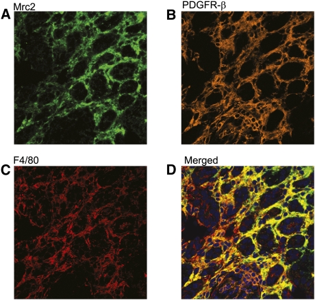
Figure 3.
Triple staining multi-photon confocal photomicrograph identifying a subset of Mrc2 interstitial myofibroblasts (PDGFR-β+) and macrophages (F4/80+) after UUO. A representative kidney section illustrates Mrc2 staining in green (detected using TSA amplified DyLight and Alexa Fluor 488-labeled secondary antibodies and the 488 laser) (A), PDGFR-β in orange (detected using TSA amplified Alexa Fluor 568-labeled secondary antibodies and the 559 laser) (B), and F4/80 in red (detected using Alexa Fluor 633-labeled secondary antibodies and the 635 laser) (C). Analysis of the merged images (D) using the Olympus FV1000MPE co-localization software program determined that 15% of PDGFR-β+ myofibroblasts and 16% of F4/80+ macrophages were Mrc2+. The photomicrographs are magnified ×400.

