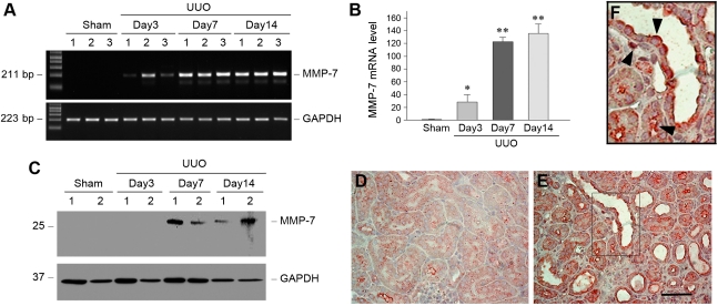
Figure 1.
MMP-7 expression is induced in a mouse model of obstructive nephropathy. (A and B) Representative RT-PCR results (A) and graphic presentation (B) show MMP-7 mRNA expression in different groups of mice as indicated. Numbers (1, 2, and 3) in A denote each individual animal in a given group. Data in B are presented as mean ± SEM of five animals (n=5). *P<0.05 and **P<0.01 versus sham controls. (C) Western blot analyses show MMP-7 protein induction in the obstructed kidney at different time points after unilateral ureter obstruction (UUO). Kidney lysates were immunoblotted with antibodies against MMP-7 and glyceraldehyde 3-phosphate dehydrogenase (GAPDH), respectively. (D and E) Immunohistochemical staining shows the localization of MMP-7 protein in obstructive nephropathy. Kidney sections from the sham (D) and unilateral ureter obstruction (E) groups 7 days after surgery were immunostained with antibody against MMP-7. Scale bar, 50 μm. (F) Enlarged image from the boxed area in E. Arrowheads indicate MMP-7–positive cells.

