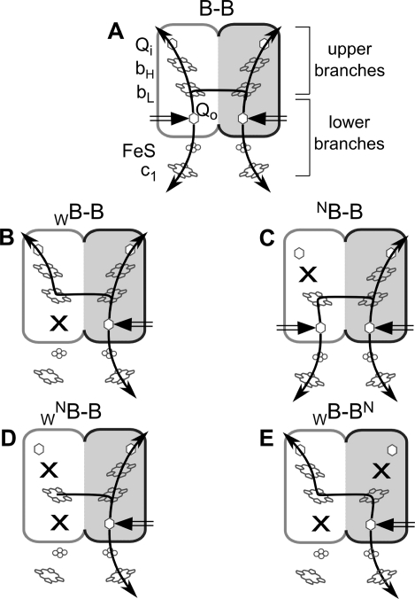
Figure 1.
Asymmetric mutation patterns in cytochrome bc1-like complexes containing fused cytochrome b subunit (B–B). The two halves of the fusion protein, each corresponding to one cytochrome b, are shown as white and gray rounded rectangles, respectively. Crosses indicate position of knockout mutations N and W, which refer to H212N and G158W point mutations in cytochrome b, respectively. Black arrows indicate functional branches. Black double arrow indicates electron entry point at the Q0 site.

