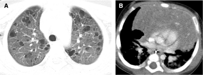
FIG. 53.
An HRCT image viewed at lung windows (A) shows numerous thin-walled cysts of various shapes in an 8-year-old with pulmonary Langerhans cell histiocytosis. A contrast-enhanced chest CT image viewed at soft tissue windows (B) shows a cavitation and calcifications within an enlarged thymus in an infant with multisystem Langerhans cell histiocytosis.

