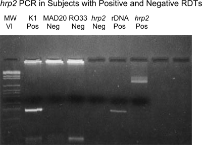
Figure 1.
hrp2 PCR in subjects with positive and negative HRP2 RDTs. Lane 1: DNA molecular weight markers VI (Roche, Indianapolis, IN). Lanes 2–6: results of PCR with DNA from a thick smear-positive subject with a negative HRP2 RDT using allotype-specific primers for the block 2 region of msp1 (lanes 2–4 show one K1 amplicon and no MAD20 or Ro33 amplicons), forward and reverse primers for hrp2 (lane 5 shows the absence of hrp2 amplicons), and species-specific primers for P. falciparum ribosomal DNA (lane 6 shows an amplicon of the expected size of 206 bp). In contrast, lane 7 provides a positive control for the hrp2 PCR based on DNA from a thick smear-positive subject with a positive HRP2 RDT (same forward and reverse primers as in lane 5 and Table 1).

