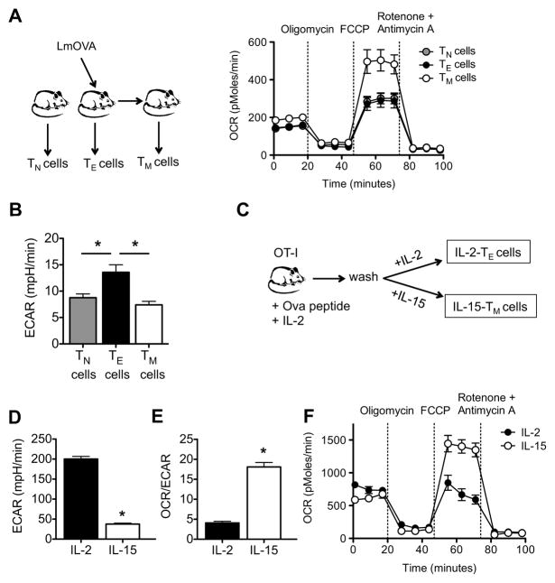
Figure 1. CD8+ TM cells have substantial mitochondrial spare respiratory capacity.
Spleens and lymph nodes were harvested from naive and LmOVA infected mice, and TN, TE, and TM cells were isolated. (A) O2 consumption rates (OCR) were measured in real time under basal conditions and in response to indicated mitochondrial inhibitors; P < 0.0001 after FCCP injection. Data are representative of 4 independent experiments. (B) Extracellular acidification rates (ECAR) were measured under basal conditions; *P < 0.01 for TE versus TN cells, and < 0.01 for TE versus TM cells. Data are representative of 2 independent experiments. (C-F) OT-I cells were activated with OVA peptide for 3 days, and subsequently cultured in either IL-2 or IL-15 to generate IL-2 TE and IL-15 TM cells, respectively. Basal extracellular acidification rate (ECAR) (D), basal OCR/ECAR ratio (E), and OCR under basal conditions and in response to indicated mitochondrial inhibitors (F) in IL-2 TE and IL-15 TM cells are shown; *P < 0.0001 (D), < 0.0001 (E), <0.0001 (F after FCCP). Data are representative of at least 3 independent experiments. Data are shown as mean ± SEM. See also Figure S1.

