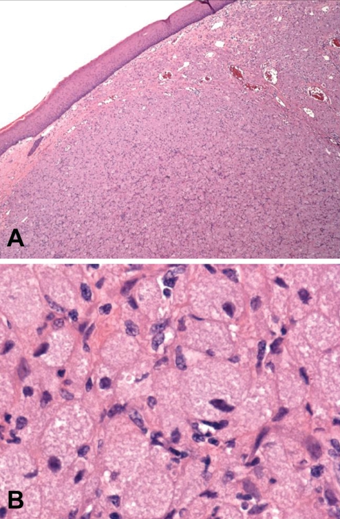
Figure 3).
A Micrograph of the congenital granular cell tumour (original magnification ×4) demonstrating the stratified squamous epithelium overlying the granular cell stroma and rich vascular supply. B Micrograph (original magnification ×60) demonstrating the polygonal cells with eosinophilic granules. Hematoxylin and eosin stain was used

