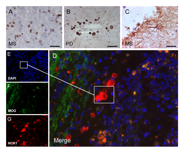
Figure 6.
NCR1+ Corpora amylacea. Using a goat polyclonal to the NCR1 C-terminus we detected numerous NCR1 round structures in WMLs (A). In Parkinson's disease tissue, near blood vessels we detected round structures bigger this time and more reminiscent of typical corpora amylacea (B). Using fluorescence microscopy these structures contained no nucleus confirming presence of NCR1 C-terminus in the membrane of corpora amylacea (D-G). NCR1 mouse monoclonal to extracellular domain was also able to detect only some corpora amylacea but not to the extent as the goat polyclonal to C-terminus and only at proximity to NCR1+ astrocytes (C). Scale bar = 10 μm (A and D), scale bar = 25 μm (B and C).

