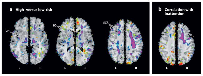
Figure 2.
Maps of local white matter volume. The statistical significance (probability values) of analyses for measures of local volumes of brain tissue, covarying for age and sex, are color-coded at each point within brain parenchyma. (a) Volumes are compared across high- and low-risk groups. Significantly reduced volumes (purple) are present in the high-risk group, primarily in bilateral dorsal frontal cortices, including the superior corona radiata (SCR) and internal capsule (IC), and in the cerebral peduncles (CP). (b) This is a map of the correlations of local volumes with measures of inattention. Regions of significant inverse correlation (purple) indicate that more inattention accompanies the reduced local volumes present in the high-risk group, primarily within the dorsal portion of the frontal white matter in the right cerebral hemisphere. L, left; R, right.

