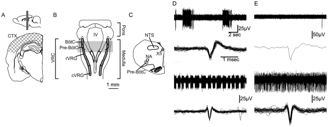
Figure 2. Location of slices for electrophysiological recordings and immunohistochemical analyses.
Spontaneous neuronal activity was recorded in cortical slices ( A ), and medullary slices ( B ) containing the rostral ventral respiratory group (rVRG), Pre Bötzinger (preBötC) and Bötzinger Complex (BötC) ( C ). Electrode arrays placed in cortical slices were centered on primary motor/somatosensory cortex (A; hatched area). Electrode arrays in medullary slices were centered on the region containing the ventral respiratory column (VRC) ( B ). The gray bar indicates the rostrocaudal extent of coronal sections used for in vitro electrophysiology and well as immunohistochemistry. During the initial equilibration, medullary neurons exhibited rhythmic bursts ( D top, bottom ), slow tonic activity ( E top ) or fast tonic activity ( E bottom ). Raw traces are shown above overlaid action potential waveforms extracted from raw data. (D, E). NA, nucleus ambiguus; IV, fourth ventricle. NTS, nucleus of the solitary tract; XII, hypoglossal nucleus; cVRG, caudal ventral respiratory group; rVRG, rostral ventral respiratory group; Pre-BötC, pre-Bötzinger complex; BötC, Bötzinger complex.

