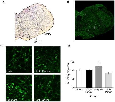
Figure 6. GABAAR ε subunit expression is increased during pregnancy in a region consistent with the rVRG and preBötC.
A, Nissl stained coronal section demonstrates anatomical location of images previously stained for semi-quantitative immunofluorescence in relation to the NA. Nuclei in the Nissl stained section were identified with reference to the rat brain atlas (Paxinos and Watson, 2004). B, Fluorescence image of the section shown in A , reacted for GABAAR ε subunit (green). The white box is placed over the region of interest imaged at 40× for quantification. C, Sample images from male, virgin female, G17 pregnant, and 30 d post-partum rats showing GABAAR ε subunit immunoreactivity. D, Quantification of integrated density of cellular label in sections from male, virgin female, G17 pregnant, and post-partum rats. * denotes significantly different than virgin female, p<0.05. Scale bar is 50 µm. NA, nucleus ambiguus; scNA, subcompact Nucleus Ambiguus.

