
Figure 1.
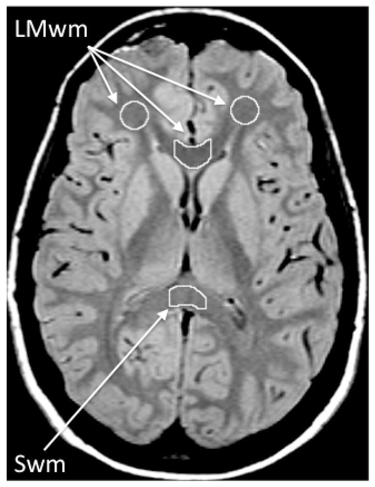
White matter regions of interest (ROI). The ROIs are depicted on an early echo (TE=20) axial MRI image that has good contrast between gray matter (light gray) and white matter (dark gray). The TE=90 (not shown) has optimal contrast between brain (appears gray) and CSF (white). Both TE=20 and TE=90 images are used to draw each ROI as this combination of slices maximizes contrast needed for optimal ROI definition. Frontal lobe white matter: The ROI is manually edited to exclude any hyperintensities or gray matter. Genu and splenium for the corpus callosum: For each of the two corpus callosum regions, a standard rectangular ROI template is first positioned on the midline, and then the anterior and posterior borders are manually edited using the contrast provided by the TE=20 and TE=90 images to exclude non-corpus callosum tissue. Lateral borders are defined by the dimensions of the rectangular template. For the genu, this positioning results in a sample consistently in the middle of the structure, which contains primarily fibers connecting the prefrontal cortices. For the splenium, this positioning samples primarily the lower half of the structure, which contains predominantly primary sensory (visual) fibers. This subject’s image was chosen as an example because the head positioning was such that frontal lobe, genu, and splenium white matter were measured on the same slices. For the majority of subjects; however, these regions are measured on different slices.
LMwm = Late-myelinating white matter (average of frontal lobe and genu of corpus callosum white matter). Swm = Splenium of corpus callosum white matter (early-myelinating region).