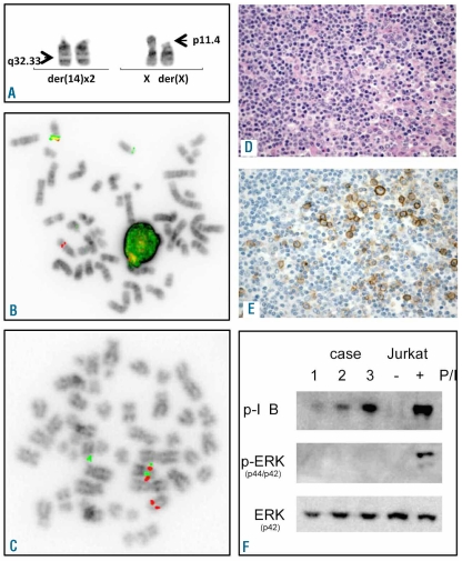
Figure 1.
Examples of cytogenetic, FISH, IHC and molecular studies. (A) Partial karyotype of case 3 showing t(X;14)(p11.4;q32.33) and an extra copy of der(14). (B and C) Examples of FISH analysis of case 3 using LSI IGH and BAC clones flanking the Xp11.4 region (RP11-204C16-SO/RP11-1174J21-SG), respectively. (D) H&E staining of case 3. (E) IHC with GPR-34 serum in case 3. (F) Immunoblot analysis of cases 1–3 with antibodies that specifically recognize phosphorylated IκBα and p42/p44 MAPK (ERK). Lysates of stimulated Jurkat cells were used as positive control. Blots were re-probed with MAPK p44 to confirm even protein loading. FISH images were acquired with a 63x/1.40 oil-immersion objective in an Axioplan 2 fluorescence microscope equipped with an Axiophot 2 camera (Carl Zeiss Microscopy, Jena, Germany) and a MetaSystems Isis imaging system (MetaSystems). H&E and IHC images were captured with a Leica DMLB microscope (Leica, Wetzlar, Germany) using a Leica PL FLUOTAR lens (40x/0.70) and a Leica DC200 camera. Images were imported directly into PowerPoint (Microsoft, Redmond, WA, USA) using the Leica DC200 camera software (version 2.51).

