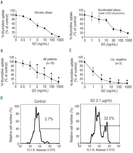
Figure 2.
Inhibition of proliferation and induction of apoptosis in CML cells by gemtuzumab/ozogamicin (GO). (A–C) Primary CML cells. (A) Individual donors. (B) All donors, n=16. (C) LSC-enriched bone marrow Lin- cells (n=3) were cultured in the presence or absence (0) of GO (1–1,000 ng/mL) at 37°C for 48 h. After incubation, 3H-thymidine uptake was measured. Results are expressed as percentage of control and represent (A) the mean±S.D. of triplicates, (B) the mean±S.D. of 16 donors, and (C) the mean±S.D. in 3 donors. (D) Mononuclear cells of a CML patient in blast phase were cultured in the presence or absence (control) of GO (0.1 μg/mL) at 37°C for 48 h. After incubation, the percentage of apoptotic cells within the CD34+/CD38− fraction was determined by combined staining with AnnexinV and cell surface markers. *P<0.05.

