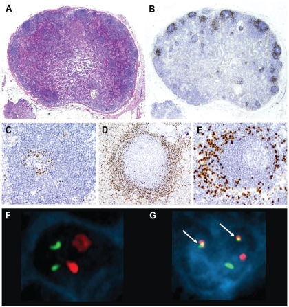
Figure 1.
In situ MCL. (A) The global architecture of the lymph node is preserved (hematoxylin-eosin stain, Olympus BX51, magnification x12.5). (B) Cyclin D1-positive cells are restricted to the mantle zones, which are not expanded (cyclin D1 stain, Olympus BX51, magnification x12.5). (C-E) “In-situ MCL” growth patterns. (C) Few cyclin D1-positive cells accumulate in the inner layer of the mantle zone (cyclin D1 stain, Olympus BX51, magnification x200). (D) Cyclin D1-positive cells that replace almost all the cells in the mantle zone, but the mantle zone is not expanded (cyclin D1 stain, Olympus BX51, magnification x100). (E) Cyclin D1-positive cells at the periphery of the mantle zone (cyclin D1 stain, Olympus BX51, magnification x200). (F, G) FISH and simultaneous cyclin D1 immunofluorescence in case 8. (F) Normal cell with negative nuclear immunofluorescence for cyclin D1; FISH using a dual-fusion probe for t(11;14) shows two red and two green signals corresponding to the presence of CCND1 and IGH genes, respectively, without rearrangement. (G) Cyclin D1 expression is shown by the blue nuclear immunofluorescence and the presence of the t(11;14) by two yellow fusion signals (arrows) in the same cell. (Olympus BX51, magnification x100, acquisition software Applied Imaging Cytovision FISH, size modified after acquisition with Adobe Photoshop).

