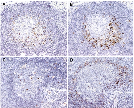
Figure 3.
(A, B) In situ MCL lesion, SOX11-positive case. The distribution of the SOX11-positive cells (A) correlates well with that of the cyclin D1-positive cells (B) (SOX11 stain (A), cyclin D1 stain (B), Olympus BX51, magnification x200). (C, D) “In situ” MCL lesion, SOX11-negative case. SOX11 is negative in the mantle zone cells (C), while cyclin D1-positive cells are seen (D). Note the follicular dendritic cells and the endothelial cells as positive internal controls for SOX11 stain (C) (SOX11 stain (C), cyclin D1 stain (D), Olympus BX51, magnification x200).

