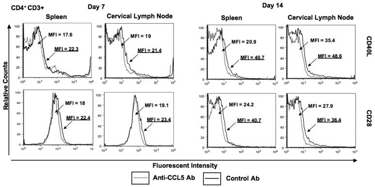
Figure 2. Flow cytometry analysis of CD28 and CD40L expression by CD4+ T cells following pneumococcal challenge.
Representative plots from three separate experiments are shown where spleen- and cervical lymph node (CLN)-derived CD4+ T cells from female F1 (C57BL/6 × BALB/c) mice, treated with control antibody (Ab, solid line) or anti-CCL5 Ab (dotted line) solutions, were isolated 7 and 14 days after intranasal challenge with Streptococcus pneumoniae strain EF3030. Mean fluorescence intensity (MFI) and fluorescence intensity histograms of CD28 and CD40L expression by CD4+ cells are illustrated and were analyzed using Flow Jo version 8.3 software. Underlined MFI values represent bacterial-challenged, anti-CCL5 antibody-treated groups.

