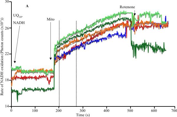
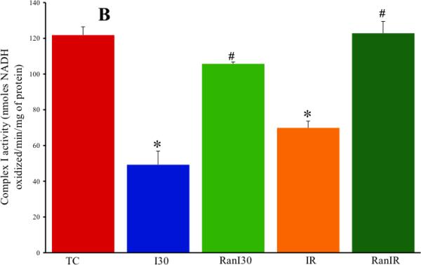
Fig. 1.


A: Representative spectrophotometric assay of mitochondrial complex I activity during cardiac ischemia reperfusion depicting the time points of addition of substrate, enzyme and the inhibitor. B: Summary data shows ischemia alone reduced the activity of the enzyme, which was restored by treatment with ranolazine. Reperfusion itself corrected the decrease in activity, but this was not as pronounced as with ranolazine (Ran) on reperfusion. Note that the activities depicted in B have been corrected for rotenone sensitivity and normalized to citrate synthase levels. * indicates p<0.05 for I30/IR vs. TC; # indicates p<0.05 for RanI30/RanIR vs. I30/IR
