
Fig. 3.
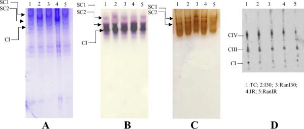
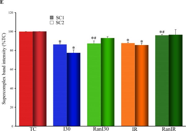
Determination of supercomplex assemblies using native PAGE. Panel A: Coomassie stained gel after native PAGE. SC1 and SC2 indicate supercomplexes composed of complexes I, III and IV, with varying copies of each complex. Components of supercomplex determined by histochemical staining for complex I (panel B), complexes III and IV (panel C) and by Western blot analysis of SC1 and SC2 against respiratory complex subunits (panel D). Summary data of supercomplex assemblies from Coomassie stained gels (panel E) shows reduction in supercomplex assemblies following ischemia and reperfusion, and restoration by ranolazine treatment. * indicates p<0.05 for I30/IR vs. TC; # indicates p<0.05 for RanI30/RanIR vs. I30/IR