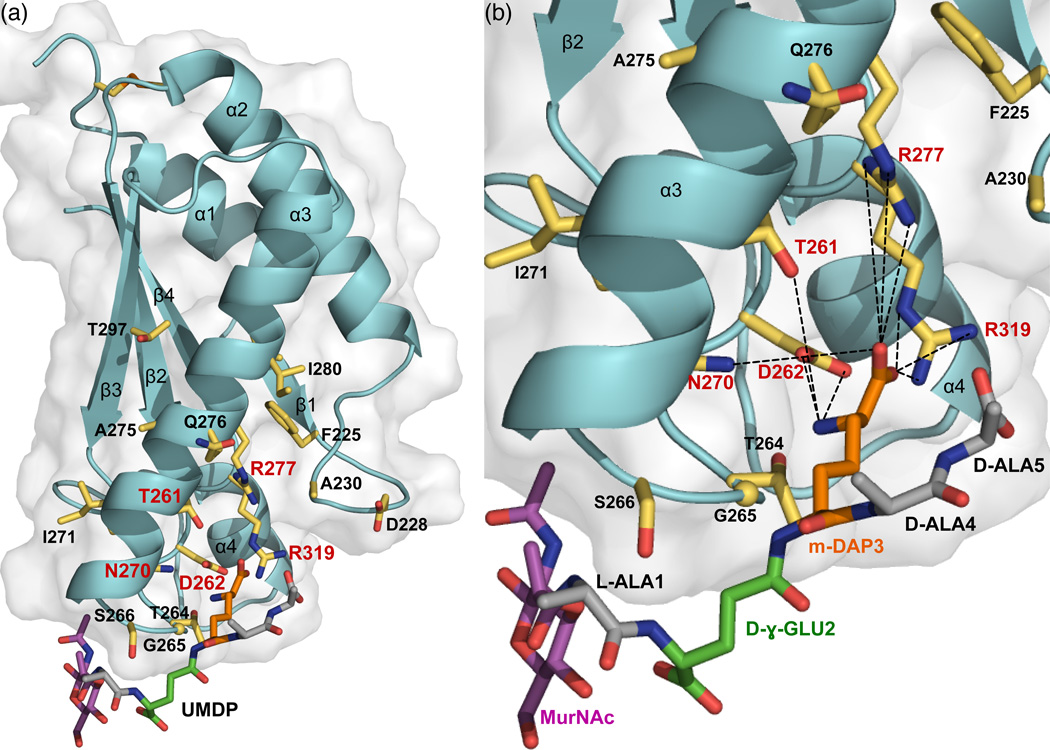
Figure 6. Peptidoglycan recognition by ArfA-c.
(a) Full and (b) close-up views of the structure of ArfA-c(D236A) associated with a structural model of the peptidoglycan peptide MDP (MurNAc–L-Ala–D-γ-Glu–m-DAP–D-Ala–D-Ala). Residues with NMR peaks most affected by UMDP (Δ≥0.03 ppm) are shown as yellow sticks. Residues that form the m-DAP recognition site are labeled in red. MDP is color coded with MurNAc in magenta, Ala in gray, D-γ-Glu in green, and m-DAP in orange.

