
Figure 4.
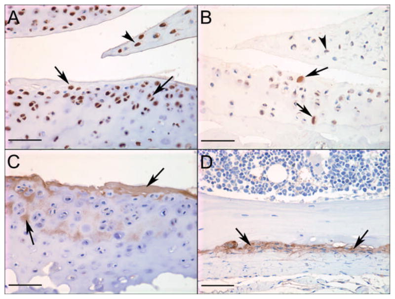
Immunohistochemical staining for selected proteins in mouse knee joints. Strong positive staining of chondrocytes in articular cartilage (arrows) and meniscus (arrowhead) for A) IL-33 in a young DMM mouse and B) CCL21 in an old sham mouse; C) positive staining for periostin in articular cartilage matrix (arrows) in a young DMM mouse and D) periosteum (arrows)in a young sham mouse; Bar = 50μm.