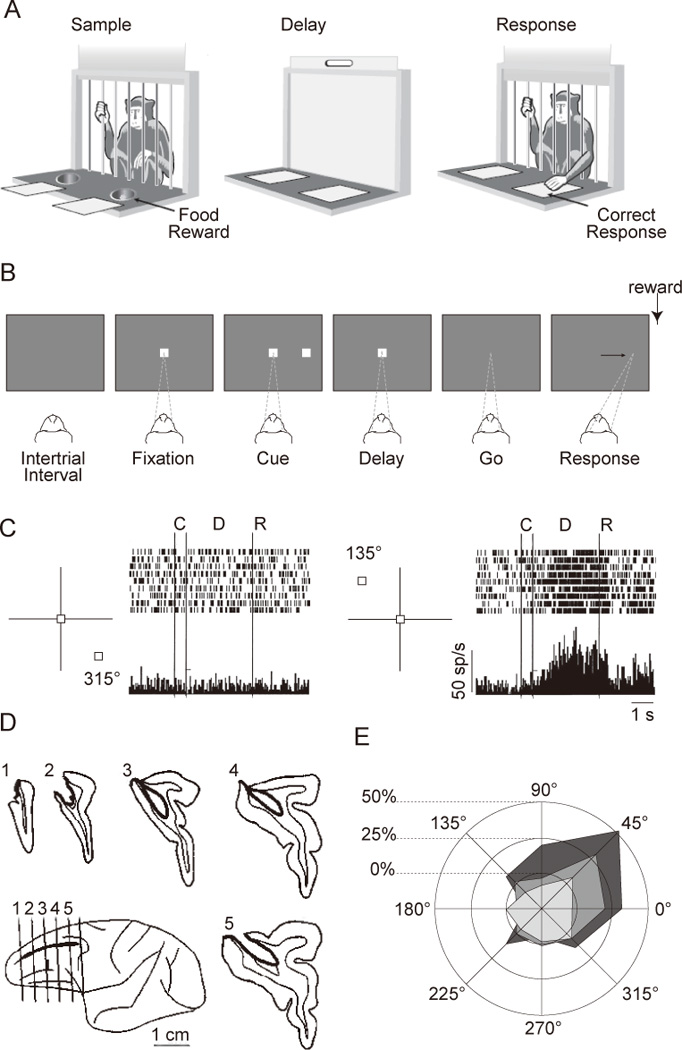
Figure 1.

Delayed-response tasks, and neuropsychology and neurophysiology using such tasks. A. Schematic drawing of a classic delayed-response task administered with the Wisconsin General Test Apparatus. In the Sample phase, the monkey observes while one of the food wells is baited with a food reward. During the Delay phase, an opaque screen is lowered, and both food wells are covered by identical objects. The Response phase is initiated by the lifting of the screen, upon which the monkey selects one of the food wells by displacing the cover in order to retrieve the reward. Illustration adapted from Curtis and D’Esposito (2004). B. Sequence of a common version of the oculomotor delayed-response (ODR) task. Each rectangle shows the screen at a time during the trial. Dashed lines and arrow show the monkey’s point of fixation. C. Activity from a DLPFC neuron located in the right hemisphere. Only trials with upper left targets (135°) and those with the opposite direction (315°) are shown. From Funahashi et al. (1989) D. Reconstruction of the lesion that gives rise to the behavioral deficit shown in panel E. From Funahashi et al. (1993). E. Changes in performance on the ODR task after unilateral lesion to the DLPFC. From Funahashi et al. (1993).
