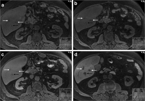
Fig. 1.
A typical case of colorectal liver metastasis. In the T1-weighted images the metastasis (black arrow) has lower signal intensity compared to the surrounding parenchyma at all phases: a, b before contrast administration, c two hours after intravenous administration of gadobenate dimeglumine and d three hours after oral ingestion of CMC-001. Some grade of enhancement of the lesion is observed after intravenous administration of gadobenate dimeglumine (c) but not after ingestion of CMC-001 (d). The white arrows indicate liver vessels

