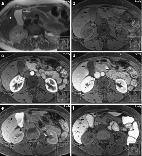
Fig. 2.
A histopathologically proven fibrotic haemangioma (arrow) being falsely classified as metastasis on both gadobenate dimeglumine and CMC-001 enhanced MRI. In a T2-HASTE image, the lesion is faintly hyperintense. In the T1-weighted images, both b before and after injection of gadobenate dimeglumine (c arterial, d portal venous and e hepatobiliary phases) as well as f 3 h after ingestion of CMC-001, the lesion has low-signal intensity, being impossible to differentiate from metastasis

