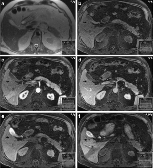
Fig. 4.
A false–negative result for gadobenate dimeglumine enhanced MRI (same patient as in Fig. 3). Both in the a T2-HASTE image as well as in the T1-weighted images after injection of gadobenate dimeglumine (c arterial, d portal venous and e hepatobiliary phases, the lesion is difficult to identify. However, in the f T1-weighted image 3 h after ingestion of CMC-001, the lesion (arrow) has clearly lower signal intensity compared to the adjacent parenchyma allowing for correct diagnosis of a metastasis

