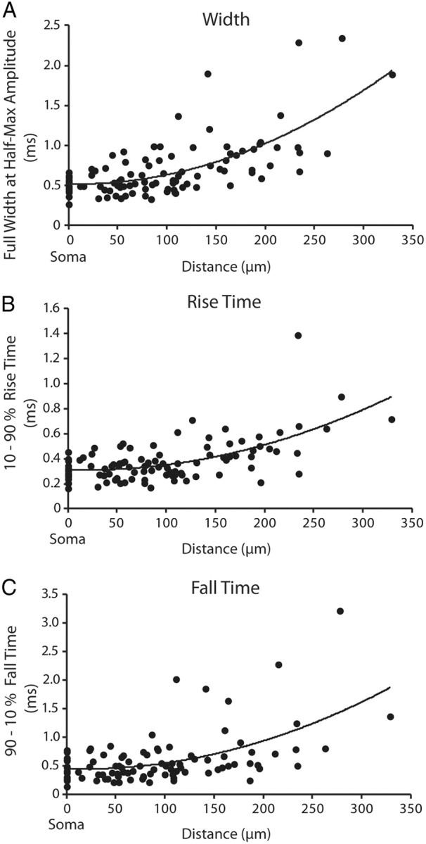
Figure 3.

Action potentials become slower and broader as they propagate to the distal dendrite. A–C, Action potential full-width at half-height (n = 30; R2 = 0.47) (A), action potential 10–90% rise time (n = 30; R2 = 0.37) (B), action potential 90–10% fall time (n = 26; R2 = 0.3) (C) plotted as a function of distance along the dendrite from the soma. Data were fit with a second-order polynomial. The somatic values (point 0) were recorded optically.
