Figure 5.
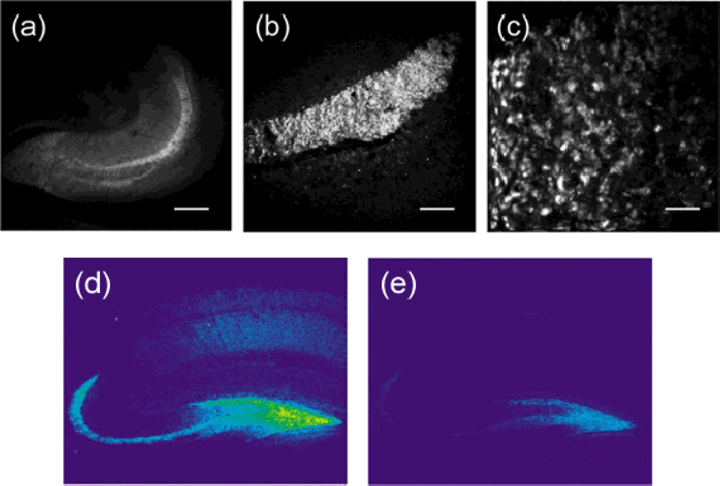
Two-photon and confocal fluorescence images of acute mouse hippocampal slices stained with ZP3. Images acquired with two-photon microscopy show (a) the entire slice section of a hippocampus, (b) the stratum lucidum layer, and (c) individual giant mossy fiber boutons. Confocal microscopy produced similar zinc-evoked fluorescence signals after ZP3 staining (d), which diminished upon addition of TPEN (e). Scale bars = (a) 800 µm, (b) 200 µm, or (c) 10 µm. Adapted with permission from (a–c) Chang et al, 2004b and (d–e) Chang et al, 2004a. Copyright (a–c) 2004 American Chemical Society and (d–e) 2004 Elsevier Science Ltd.

