Figure 1.
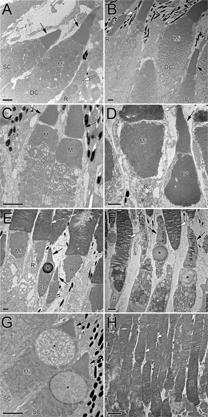
Electron micrographs of vertebrate photoreceptors illustrating diverse outer segment tapers and ellipsoid morphologies. (A) Goldfish single cone (SC) and rod (R) flanking one member of a double cone (DC). The cone ellipsoids are packed with mitochondria (Mi). Cone outer segments (black arrows) taper, whereas those of the rods do not (white arrow; only a portion of the rod outer segment is visible). (B) Single cone, double cone, and rod of coho salmon. In this species, there is a clear gradient in the size of cone mitochondria from smaller, at the level of the myoid, to larger, at the level of the ellipsoid. (C) Double cone from a mummichog killifish showing megamitochondria (M) associated with the ellipsoid of each double cone member. This species also has ellipsosomes, which arise from megamitochondria as the cristae disappear. (D) Rod and single cone from a bullfrog. The rod mitochondria are long and compacted; the single cone exhibits an ellipsosome-like structure (E*) in the ellipsoid. (E) Two single cones among rods in the bullfrog retina; one of the cones contains an oil droplet (*). Note the large difference in size between rods and cones. (F) Single cones and rods from a Canada goose. The single cones show different types of oil droplets. As in the frog, elongated mitochondria pack rod inner segments, and the mean diameter of cone ellipsoids (entrance aperture) is similar to that of rods. (G) Single cones of the red-eared slider turtle showing large oil droplets and pronounced cone taper. (H) Rods of the mouse retina. The cones in this and similar nocturnal species are hard to identify without molecular markers. Bars, 2 µm.

