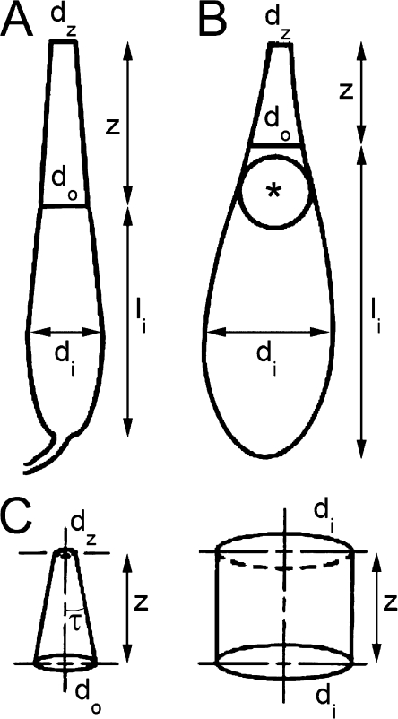
Figure 2.
Drawings of single cones from fresh retinal preparations illustrating the morphological parameters measured as well as the taper angle, τ. The cellular dimensions were obtained from video images recorded via a microscope equipped with a calibrated infrared-sensitive video system. (A) Single cone from blue gill sunfish. (B) Single cone from leopard frog. (C) Cone outer segment (left) from B and an idealized representation of that of the optically equivalent rod (right). The equivalency is based on the assumption that both cells have equal entrance aperture with diameter di and that the cone ellipsoid funnels the incident flux to the outer segment without loss. The cellular dimensions (in µm) for these cones were as follows: (A) for the blue gill sunfish, di = 8.3, do = 5.0, dz = 2.9, z = 18, and the inner segment length, li = 25.2; (B) for the leopard frog, di = 7.2, do = 2.8, dz = 1.3, z = 6.3, and li = 17.5. The parameter z, in these two cases, equals the outer segment length, and dz is the diameter at the tip of the outer segment. The asterisk in B depicts an oil droplet.

