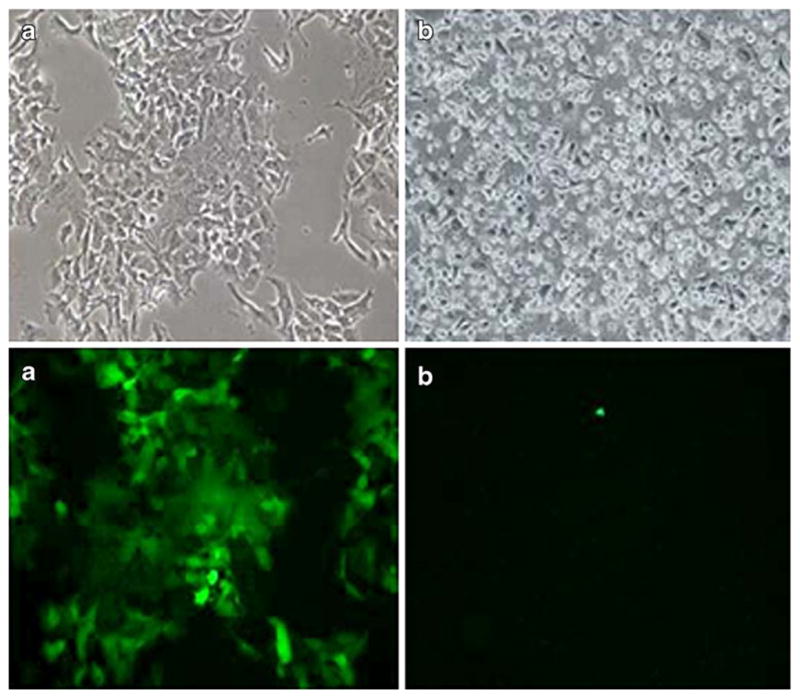
Fig. 2.

Green fluorescent protein (GFP) expression of MKN74 (a) and AGS (b) cell lines 5 days after infection with NDV(F3aa)-GFP. Abundant GFP expression was observed in the MKN74 (a) cell lines. AGS (b) displayed minimal GFP expression (×100 magnification)
