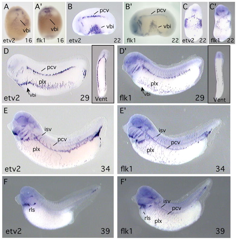
Fig. 2. etv2 is expressed in hematopoietic and endothelial precursor cells in the Xenopus embryo.
Transcripts of etv2 (A–F) and flk1 (A′-F′) were detected by in situ hybridization. flk1 is an established marker of the hematopoietic and endothelial lineages. Developmental stage of the embryos is indicated at the lower right of each panel. Embryos shown in A, A′, C, C′ and insets in D, D′ are ventral views. All other views are lateral with anterior to the left. Abbreviations: vbi, ventral blood island; pcv, posterior cardinal vein; plx, vascular plexus; isv, intersegmental vessel; rls, rostral lymphatic sac.

